While most eyes stay glued to the brain in the hunt for Alzheimer’s treatments, another organ quietly steps into the spotlight.
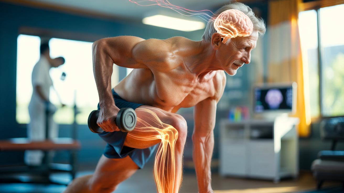
New research suggests that the path to protecting memory might begin far from neurons and synapses, in an organ usually associated with dumbbells and marathons: skeletal muscle.
Muscles talking to the brain: a hidden communication channel
For years, muscle was treated as little more than a motor: it contracts, moves bones, burns energy. End of story. That picture is now outdated. Scientists increasingly view muscle as an endocrine organ that releases chemical messengers into the bloodstream.
These messengers, called myokines, are proteins secreted during and after muscle contraction. Once released, they travel through the circulation and can reach distant organs, including the brain. Some myokines influence inflammation, others energy use, and a few appear to shape cognition.
Among them, one molecule has drawn special attention: cathepsin B. Levels of this protein rise after physical activity, and previous studies linked it to improved learning and memory in both animals and humans.
Cathepsin B, released by active muscles, has been associated with brain plasticity and the birth of new neurons in memory-related regions.
Scientists began to wonder whether this “muscle signal” could help a brain under attack from a neurodegenerative disease. Instead of hitting the usual targets in Alzheimer’s – amyloid plaques and tau tangles – they asked a different question: could strengthening the conversation between muscle and brain make the brain more resilient?
A muscle‑based therapy tested in an Alzheimer’s mouse model
To test that idea, researchers turned to mice genetically engineered to develop features resembling Alzheimer’s disease. These animals usually show memory deficits, reduced learning abilities and a drop in the production of new neurons in the hippocampus, a crucial hub for memory formation.
The team used a viral vector – a harmless, engineered virus – designed to boost production of cathepsin B specifically in skeletal muscle. The brain itself was not directly manipulated. The goal was simple: get the muscles to talk louder to the brain.
Six months later, the treated mice looked surprisingly different from their untreated counterparts during memory tests. Their spatial memory remained stable, and their learning performance approached that of healthy, non-Alzheimer’s mice.
➡️ Abandoned in adulthood, this habit could quietly extend your wellbeing
➡️ Caffeine Becomes A Molecular Switch Envisioned For Treatment
➡️ Gut microbiome and daily flatulence: unexpected numbers
➡️ Unexpected find: thousands of nests spotted beneath Antarctic ice
➡️ Because of our lifestyle, osteoarthritis is surging among young adults worldwide
➡️ The “brain‑eating” amoeba shrugs off chlorine and slips into our water systems
➡️ Loose eyelid syndrome can reveal a hidden sleep disorder
➡️ An Oat-Based Diet Cuts Cholesterol In Just Two Days
Despite carrying genetic risk and typical brain lesions, mice with boosted muscle cathepsin B maintained stronger learning and memory abilities over time.
When the researchers examined the animals’ brains, they found that neurogenesis – the formation of new neurons – in the hippocampus was restored. Under usual Alzheimer-like conditions in mice, this process drops sharply. Here, it bounced back.
Proteins in the brain, muscle and blood also showed a shift. Their overall patterns resembled those seen in healthy animals, suggesting that the muscle intervention had rebalanced broader biological pathways rather than acting at a single, isolated target.
Alzheimer’s markers stay, memory improves
One of the most striking findings came when the scientists checked the classical hallmarks of Alzheimer’s in these mice. Despite the cognitive improvements, amyloid deposits and signs of brain inflammation remained.
The lesions were still there, yet the animals remembered better.
The study suggests that memory can be preserved without wiping out amyloid plaques, by boosting the brain’s capacity to adapt and function despite damage.
Cathepsin B seemed to trigger a cascade of protein changes related to cell growth, synaptic plasticity and neurogenesis. In other words, instead of scrubbing the brain clean, the signal from muscle helped the brain reorganise and keep working under stress.
The story comes with a twist. In healthy mice without Alzheimer-like pathology, artificially raising cathepsin B too much produced the opposite effect: memory problems appeared. That contrast hints at a context-dependent action. The intervention supports a vulnerable brain but does not act as a general cognitive enhancer.
Why muscles matter for an ageing brain
This line of work bolsters a broader idea: Alzheimer’s is not solely a brain disease. The rest of the body, from fat tissue to the immune system and muscles, shapes how the disease unfolds.
Muscle sits at an interesting crossroads. Ageing often leads to sarcopenia, the gradual loss of muscle mass and strength. At the same time, dementia risk climbs. Researchers suspect that reduced muscle activity means fewer beneficial myokines reaching the brain, leaving neurons with less support.
Studies in humans have already linked regular physical exercise with better cognition and lower dementia risk. The new data help explain one possible biological chain connecting squats in the gym to sharper memory decades later.
- Aerobic exercise raises production of several myokines, including cathepsin B and irisin.
- These molecules influence brain plasticity and blood flow.
- Improved vascular health and synaptic function may delay or reduce cognitive decline.
From lab strategy to future treatments
The mouse work published in 2025 in the journal Aging Cell is still far from a therapy for humans. Gene delivery into muscle using viral vectors raises safety and ethical questions, and animal models do not fully mimic human Alzheimer’s.
Yet the concept behind the study opens up several avenues for future research:
- Drugs that gently boost specific myokines in muscle.
- Exercise programmes tailored to maximise protective signals towards the brain.
- Biomarkers in blood that reflect the quality of muscle–brain communication.
Instead of directly attacking plaque formation in the brain, future interventions might work by reinforcing the body’s built-in defence networks. Muscle could become a target organ for dementia prevention programmes, alongside more traditional cardiovascular and metabolic measures.
Key concepts: myokines, neurogenesis and brain plasticity
Several scientific notions sit at the heart of this research. Understanding them helps clarify why muscle signals might matter for memory.
| Term | What it means | Why it matters for Alzheimer’s |
|---|---|---|
| Myokines | Proteins released by muscles into the bloodstream during contraction. | Some myokines influence inflammation, energy balance and brain function. |
| Neurogenesis | Process by which new neurons are created from stem cells, especially in the hippocampus. | Reduced neurogenesis is linked to memory problems; restoring it could support learning. |
| Synaptic plasticity | Ability of connections between neurons to strengthen or weaken in response to activity. | Crucial for encoding memories and adapting to damage. |
| Endocrine organ | An organ that secretes hormones or signalling molecules into the blood. | Viewing muscle as endocrine shifts it from passive tissue to active regulator. |
What this could mean for everyday life
While no one should expect a workout to “cure” Alzheimer’s, the muscle–brain link adds weight to current advice around staying active, especially from midlife onwards. The benefits likely stack up over time, touching both vascular health and direct brain signalling.
Consider two ageing scenarios. In the first, an older adult remains mostly sedentary, gradually losing muscle mass, balance and endurance. Myokine output shrinks, blood vessels stiffen and small brain injuries accumulate faster. In the second, the same person keeps up regular brisk walks, light resistance training and social activity. Their muscles stay more active, feeding the brain a richer mix of protective signals. The underlying genetic risk for Alzheimer’s may be the same, but the trajectory of decline could be very different.
There are also risks to keep in mind. Overstimulating certain pathways, as seen with excessive cathepsin B in healthy mice, may backfire. Future therapies will likely depend on careful dosing and on identifying who stands to benefit most – for instance, people already showing early cognitive changes or carrying high-risk genetic variants.
For now, the core message is less about futuristic gene tools and more about reframing how we think of muscle. Each contraction is not just mechanical work; it is also a chemical broadcast. That broadcast can reach the brain and, under the right conditions, help it hold onto memories a little longer in the face of disease.